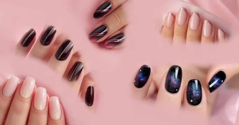
Nails are complex, keratin-based structures that play an essential role in protecting the extremities of the fingers and toes. They are made primarily of keratin, a fibrous protein that is also found in hair and the outer layer of skin. The anatomy of the nail reveals intricate parts, each contributing uniquely to the formation, growth, and function of nails.
At the core of nail formation is the nail matrix (or germinal matrix). This is the living tissue located beneath the skin at the base of the nail, under the cuticle. The matrix is crucial because it produces new cells that harden and compact into the visible nail plate as they move outward. The matrix contains nerves, blood vessels, and lymph, which help supply nutrients needed for nail growth. The size and health of the matrix determine the thickness and width of the nails. An interesting part of the matrix visible through the nail plate is the lunula, the half-moon shaped whitish area at the nail’s base. It usually appears more prominently on the thumbs than other fingers. The shape of the nail plate is also influenced by the underlying bone structure of the fingertip.
The nail plate is what is commonly recognized as the nail — the hard, translucent structure visible on the finger or toe. It consists of several layers of dead, densely packed keratinized cells that provide strength and protection. Unlike bone, the nail plate contains no blood vessels or nerves, which is why damage or pressure on the plate itself is less painful compared to the surrounding tissue. The plate is tightly attached to the nail bed — the skin beneath it — which is rich in blood vessels, giving nails their pinkish hue. The nail bed supports the nail plate and nourishes it as it grows.
Surrounding the nail plate are several protective structures. The nail folds are the skin that overlaps the edges of the nail, helping anchor and protect the nail from injury and infection. The cuticle (or eponychium) is a thin layer of living skin that grows from the nail fold to the base of the nail plate and helps seal the area so that pathogens cannot enter the nail matrix. Removing the cuticle can expose the matrix to infections and complications.
At the distal end, the nail plate extends beyond the fingertip as the free edge, which can be trimmed. Underneath this free edge is the hyponychium, a thickened layer of skin that forms a seal between the nail plate and fingertip, preventing dirt and microbes from entering and protecting the delicate tissue underneath. Near the free edge is also the onychodermal band, a translucent seal visible as a slight color change on the nail, which serves as an additional protective barrier.
Functionally, nails serve several important roles:
- They protect the fingertips and toes from mechanical damage. The fingertips are highly sensitive and exposed, so the hard nails shield them against injuries.
- Nails enhance sensory perception by providing a firm backing to the fingertips. This backing allows fingertips to better detect fine textures and small objects through enhanced tactile feedback.
- They assist in manipulating small objects such as picking, scratching, or opening items.
- Nails contribute to the aesthetic and functional integrity of the hands and feet, influencing both appearance and the ability to perform daily tasks.
Nail health is also an indicator of overall health. Changes in nail color, thickness, texture, or growth rate can signal infections, nutritional deficiencies, or systemic diseases.
In summary, nails are sophisticated structures composed mainly of keratinized cells produced by the nail matrix. They work in conjunction with surrounding skin tissues to protect the digits, enhance tactile ability, and aid in grasping and manipulation. The intricate anatomy, including the nail plate, matrix, bed, folds, cuticle, and hyponychium, plays specialized roles to maintain nail growth, protection, and overall function.


